Molecular diagnostics of cancer have highlighted the significance of driver genetic alterations in tumor growth and metastasis. Although genetic variations offer valuable diagnostic and therapeutic insights for cancer patients, detecting mutations in circulating cell-free DNA (cfDNA) proves to be a major challenge due to their typically low levels. One of the primary obstacles in the sensitive detection of low-level altered DNA is the intense background noise arising from an abundance of wild-type (WT) DNA strands.
Currently, DNA mutation detection methods fall into two categories: direct detection methods and enrichment-based methods. While direct detection methods like qPCR, droplet digital PCR (ddPCR), and sequencing have detection limits greater than 0.1%, enrichment techniques face challenges in clinical applications due to complex probe design, temperature sensitivity, cost of auxiliary instruments/reagents, and limited general applicability.
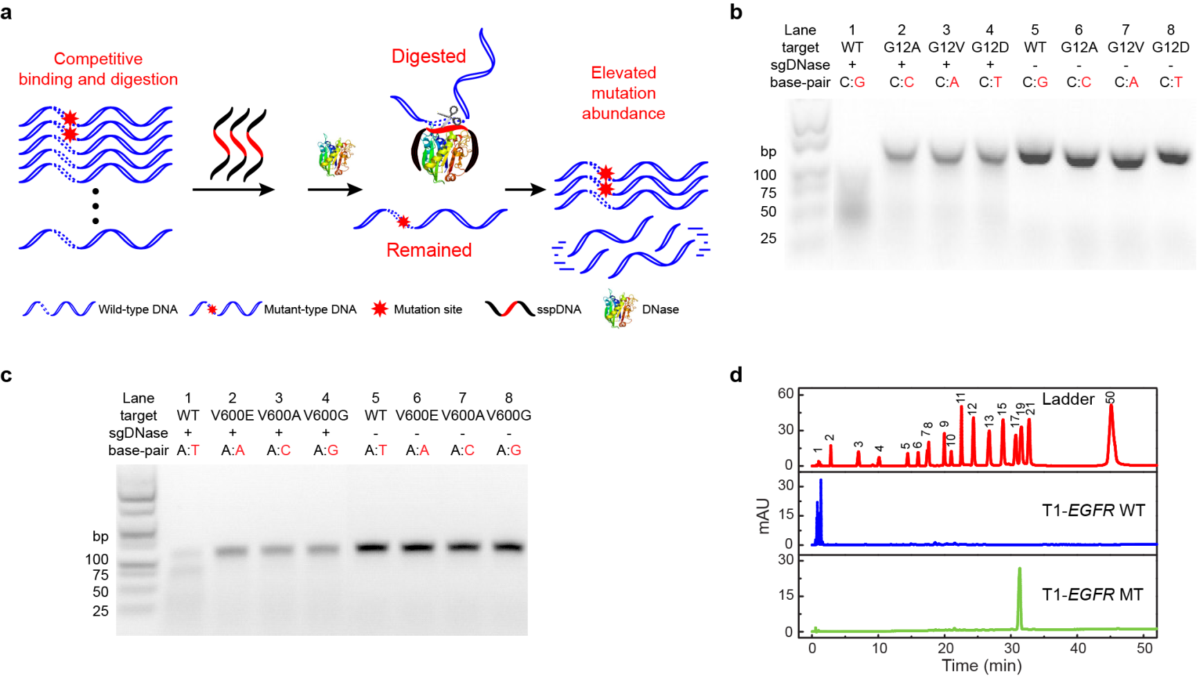
Recently, the research team led by Professor Meiping Zhao at the College of Chemistry and Molecular Engineering, Peking University (PKU-CCME), published a research paper titled "Detection of low-frequency mutations in clinical samples by increasing mutation abundance via the excision of wild-type sequences" in Nature Biomedical Engineering. Their major breakthrough involved developing a precise DNA excision tool based on a competitive DNA-binding-and-digestion mechanism. This mechanism is facilitated by deoxyribonuclease I (DNase) guided by single-stranded phosphorothioated DNA (sgDNase), enabling the removal of WT DNA strands (Fig. 1a). By guiding DNase with appropriate sspDNA, they achieved single-nucleotide resolution, surpassing their previously reported DNase/sspDNA complexes(Chemical Science, 2016, 7, 2051).They first validated the sequence specificity of sgDNase by fluorescent probe method, high-performance liquid chromatography analysis and gel electrophoresis analysis respectively. As shown in Fig. 1b–d, under the guidance of sspDNA with a sequence fully complementary to the WT DNA sequence, sgDNase could rapidly digest WT DNA strands with high selectivity. In contrast, the majority of the mutant-type DNA strands (MT) were retained. Moreover, the results demonstrate the discrimination effect of sgDNase on different types of mismatches across various genes.

Fig.1 Proof of concept: phosphorothioated DNA guided deoxyribonuclease I (sgDNase).(a)Illustration of the sgDNase system via a competitive binding and digestion mechanism.(b–d) Determination of the single-base differentiation ability of sgDNase between fully matched wild-type strand and mismatched mutant-type strands with different types of mismatched bases using(b, c) agarose gel electrophoresis analysis and (d) HPLC of the digestion products of (b) KRAS G12 targets, (c) BRAF V600 targets and (d) EGFR L858 targets.
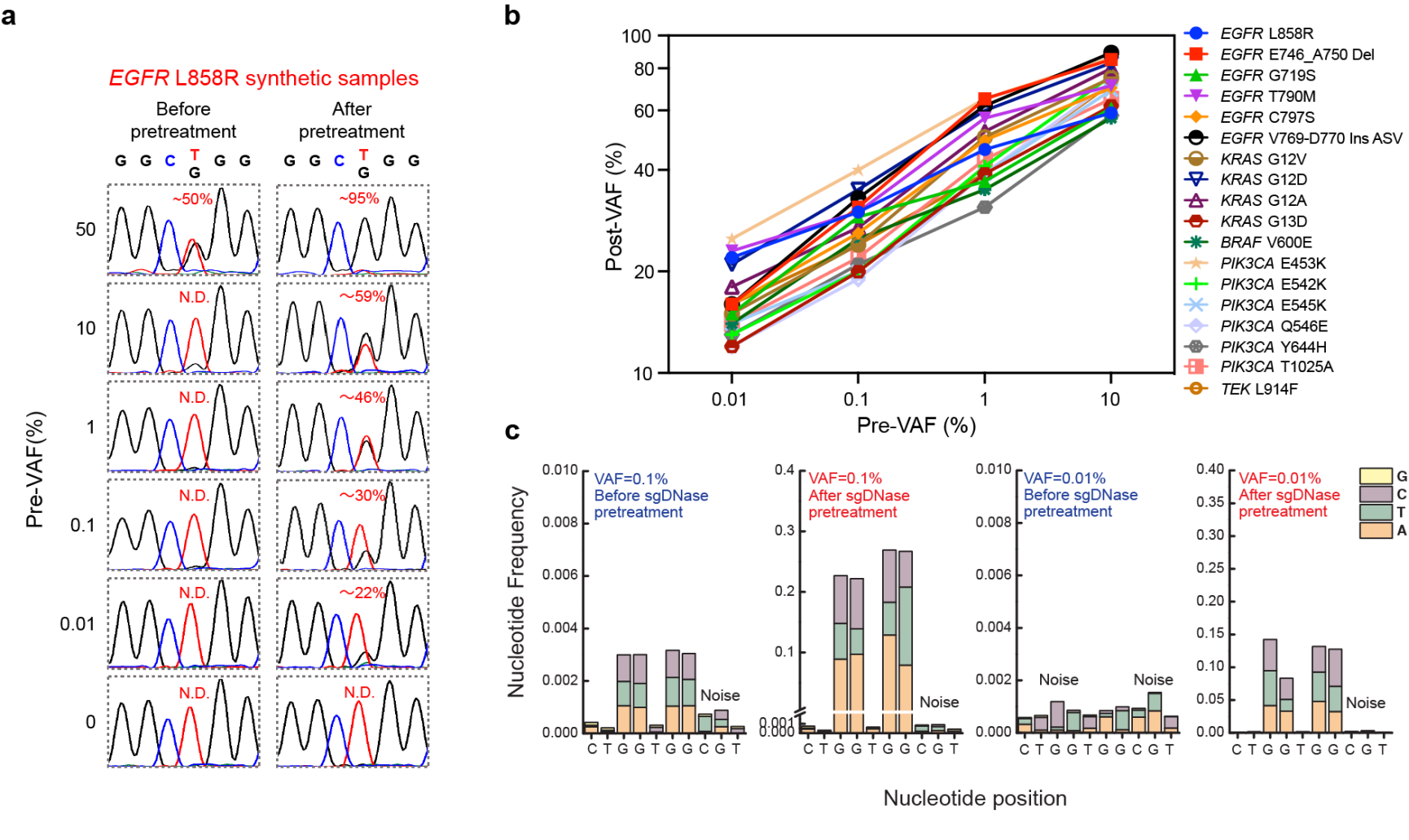
The effectiveness of sgDNase was demonstrated in enriching low-level mutant DNA strands, particularly the EGFR L858R mutation. By pretreating synthetic samples, they significantly increased the variant allele frequency (VAF) from 0.01% to 22% (Fig. 2a). Moreover, sgDNase was successful in enriching 18 different mutations with initial VAFs ranging from 0.01% to 10% (Fig. 2b). Furthermore, the sgDNase system enabled the simultaneous enrichment of different mutation types within mutational hotspots, such as KRAS G12 and G13, facilitating the quick identification of potential rare and unknown mutations (Fig. 2c).

Fig.2 Enrichment of single and multiplex hotspot mutations by sgDNase pretreatment. (a) Comparison of Sanger sequencing chromatograms of synthetic EGFR L858R samples with different pre-VAFs (0%–50%) before and after sgDNase pretreatment. (b) Post-VAFs (measured by Sanger sequencing) of 18 clinically relevant target mutations with pre-VAFs varying from 0.01% to 10% after single-plexed enrichment by sgDNase. (c) Next-generation sequencing (NGS) measurement results of the abundance of KRAS G12G13 mutations in the sample with a pre-VAF at 0.01% before and after sgDNase pretreatment. Variant to noise plots derived from sequencing data are depicted.
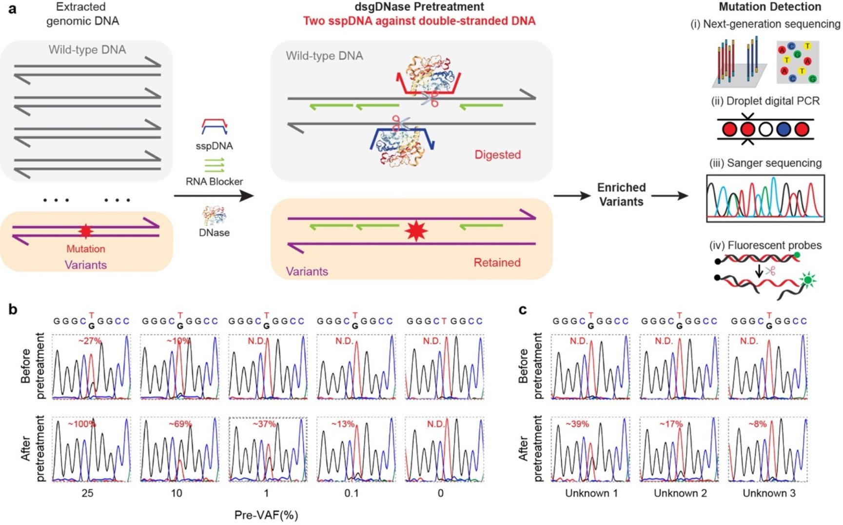
They extended the application of sgDNase to directly pretreat double-stranded genomic DNA using the double phosphorothioated DNA guided deoxyribonuclease I (dsgDNase) system. This approach significantly increased VAFs for various mutations, enabling their detection using multiple mutation detection methods (Fig. 3a). Notably, the sgDNase pretreatment allowed for accurate identification of ultra-low-abundance mutations in clinical biopsy samples (Fig.3b and 3c).

Fig.3 Direct pretreatment of genomic DNA by double sspDNA guided DNase (dsgDNase). (a) Schematic illustration of direct enrichment of mutations in double-stranded genomic DNA by double phosphorothioated DNA guided deoxyribonuclease I (dsgDNase) system. (b) Comparison of the Sanger sequencing results of genomic DNA with EGFR L858R mutation at different pre-VAFs (0%–25%) before and after dsgDNase pretreatment. (c) Sanger sequencing chromatograms of three unknown genomic DNA samples with EGFR L858R mutation before and after dsgDNase pretreatment.
The sgDNase assay has been applied to more than 70 clinical samples and totally 16 positive samples were identified. As shown in Fig. 4a,b, without sgDNase pretreatment, only two tissue samples (Patients P2 and P5) were observed to be EGFR L858R mutation positive, while no positive blood samples were identified. After sgDNase pretreatment, one more tissue sample (Patient P7) and two blood samples (Patients P2 and P10) were identified as L858R mutation positive (Fig.4a). The positive samples were further validated by NGS. Elevated VAFs were achievedin clinical samples, regardless of sample types (blood, tissue) and disease types (lung cancer, colorectal cancer and venous malformations) after sgDNase pretreatment. Thes gDNase exhibited a clinical sensitivity of 94.74% and a clinical specificity of 98.58%.

Fig.4 Detection of ultralow-abundance mutations in clinical biopsy samples with sgDNase pretreatment. (a) sgDNase applied to sixteen cfDNA and genomic DNA samples from eight NSCLC patients (P1-P8), four colorectal cancer patients (P9-P12) and standard reference DNA (SRD). P, T and B in the sample name represent patient, tissue sample (genomic DNA) and blood sample (cfDNA) respectively. (b) Comparison of Sanger sequencing chromatograms of the EGFR L858R mutation in gDNA (tissue) and cfDNA (blood) samples from an NSCLC patient and a colorectal cancer patient before and after sgDNase pretreatment. (c) Summary of the post-VAFs of cancer patients and negative gDNA control after sgDNase pretreatment (measured by Sanger sequencing). P value = 1.4E-76, from two-tailed Welch’s unpaired t-tests.
The high-resolution capability, universality, ease of implementation, and robust enrichment capability of sgDNase hold significant potential for continuous mutation status assessment and precise cancer therapy. Additionally, sgDNase can be employed to selectively digest unwanted DNA sequences for various detection purposes, further enhancing its versatility and applicability.
The development of the phosphorothioated DNA-guided endonuclease excision tool represents a groundbreaking advancement in DNA mutation detection. Its ability to remove WT DNA background and enrich low-frequency mutations opens up new possibilities for accurate diagnosis and targeted cancer therapy. The simplicity, efficiency, and broad applicability of this method make it a promising asset in the field of molecular diagnostics.
The joint first authors of the article are WeiChen (a Ph.D. graduate), HaiqiXu (a B.S. graduate), and Shenbin Dai (a Ph.D. candidate) from the College of Chemistry and Molecular Engineering, Peking University. Prof. Meiping Zhao from the College of Chemistry and Molecular Engineering, Peking University, Prof. Xianjin Xiao (a 2016 Ph.D. alumnus of PKU-CCME) and Prof. Tongbo Wu (a 2017 Ph.D. alumnus of PKU-CCME), both from Tongji Medical College, Huazhong University of Science and Technology, are the co-corresponding authors. The project was funded by the National Natural Science Foundation of China and the Beijing National Laboratory for Molecular Sciences.
Link to the original article: https://www.nature.com/articles/s41551-023-01072-8
Full-text access link:https://rdcu.be/dhUlL